Anatomy Of Chest And Abdomen / Anatomy of human abdominal muscles with labels — text ... - But with the use of smart technology, you can learn faster and master abdomen anatomy in no time!
Anatomy Of Chest And Abdomen / Anatomy of human abdominal muscles with labels — text ... - But with the use of smart technology, you can learn faster and master abdomen anatomy in no time!. Abdominal computed tomography (ct) is a type of medical imaging procedure used to diagnose and monitor internal stomach issues, like cancer, bowel obstruction, and abdominal this photo gallery presents the anatomy of the abdomen by means of ct (axial, coronal, and sagittal reconstructions). 14 chest selected topics lungs and airways vascular anatomy nodal stations gi abdomen and pelvis gi hepatic segmental anatomy vascular anatomy nodal groups. But with the use of smart technology, you can learn faster and master abdomen anatomy in no time! With skeletal, chest and abdomen pattern differentials dennis marchiori (auth.). Thorax, heart, abdomen clinical imaging.
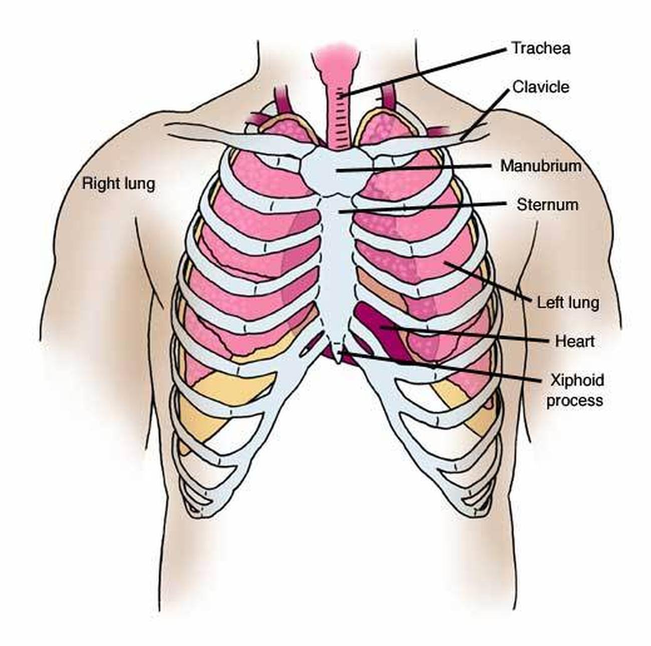
They serve to protect the internal organs of the thorax while allowing the lungs to expand during respiration. The diaphragm forms the upper surface of. First the heart figure is evaluated, followed by mediastinum and hili. Diseases affecting any of these organs could result in abdominal pain. Subsequently the lungs, lungborders and finally the chest wall and abdomen.
Pictures Of Chest from healthiack.com Radiology basics of abdominal ct anatomy with annotated coronal images and scrollable axial images to help medical students and junior doctors learning anatomy. 1900 human anatomy print chest and abdominal organs. Its upper boundary is the diaphragm, a sheet of muscle and connective tissue that separates it the abdominal organs are supported and protected by the bones of the pelvis and ribcage and are covered by the greater omentum, a fold of peritoneum. The principle parts of the human body are the head, the trunk and the limbs (extremities). The epidermis is the outermost layer that provides a protective, waterproof seal over the body. Human anatomy of female chest and abdomen stock photo. Anatomy of the chest and the lungs: Pathology of the heart, mediastinum, lungs and pleura.
The abdomen (colloquially called the belly, tummy, midriff or stomach) is the part of the body between the thorax (chest) and pelvis, in humans and in other vertebrates.
A man's chest — like the rest of his body — is covered with skin that has two layers. The mdct anatomy of the chest, abdomen, and pelvis is presented in three different parts. A curated list of anatomical features in the human abdomen and relevant open datasets. Use the mouse scroll wheel to move the images up and down alternatively use the tiny arrows (>>) on both side of the image to move the images. 14 chest selected topics lungs and airways vascular anatomy nodal stations gi abdomen and pelvis gi hepatic segmental anatomy vascular anatomy nodal groups. Thus, the right side of the image is the patient's left. Abdominal computed tomography (ct) is a type of medical imaging procedure used to diagnose and monitor internal stomach issues, like cancer, bowel obstruction, and abdominal this photo gallery presents the anatomy of the abdomen by means of ct (axial, coronal, and sagittal reconstructions). Let's take a close look at this very important part of our anatomy and thus improve our understanding of causes of abdominal pain. The abdomen (colloquially called the belly, tummy, midriff or stomach) is the part of the body between the thorax (chest) and pelvis, in humans and in other vertebrates. Pathology of the heart, mediastinum, lungs and pleura. 1900 human anatomy print chest and abdominal organs. The abdomen (commonly called the belly) is the body space between the thorax (chest) and pelvis. It can help you understand our world more detailed and specific.
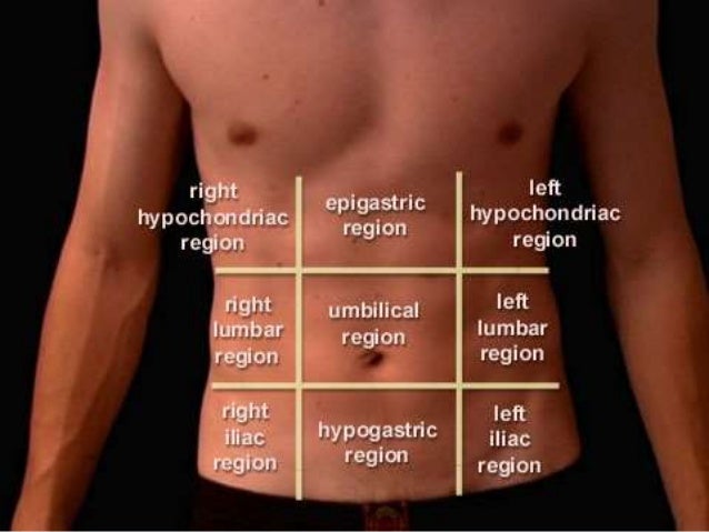
Its upper boundary is the diaphragm, a sheet of muscle and connective tissue that separates it the abdominal organs are supported and protected by the bones of the pelvis and ribcage and are covered by the greater omentum, a fold of peritoneum. The median plane is that which follows the linea alba and extends from the xiphoid process to the. They are separated by theoretical anatomical lines that can be traced on the abdomen using certain anatomical landmarks. The human abdomen is that part in the front of our body between the chest and the waist line. This mri abdomen axial cross sectional anatomy tool is absolutely free to use.
Anatomy of abdomen and regions of trunk from image.slidesharecdn.com The mdct anatomy of the chest, abdomen, and pelvis is presented in three different parts. Diseases affecting any of these organs could result in abdominal pain. Radiology basics of abdominal ct anatomy with annotated coronal images and scrollable axial images to help medical students and junior doctors learning anatomy. Human anatomy of female chest and abdomen stock photo. Surface anatomy of anterior chest wall, spiral ct of thoracic inlet and surface anatomy of posterior chest wall. Superior part of trunk including pector… This mri abdomen axial cross sectional anatomy tool is absolutely free to use. Anatomy is the amazing science.
But with the use of smart technology, you can learn faster and master abdomen anatomy in no time!
With skeletal, chest and abdomen pattern differentials dennis marchiori (auth.). They serve to protect the internal organs of the thorax while allowing the lungs to expand during respiration. The median plane is that which follows the linea alba and extends from the xiphoid process to the. Learn about chest and abdomen anatomy with free interactive flashcards. Ribs are long curved bones which form the rib cage surrounding the chest. Let's take a close look at this very important part of our anatomy and thus improve our understanding of causes of abdominal pain. Thorax, heart, abdomen clinical imaging. Radiology basics of abdominal ct anatomy with annotated coronal images and scrollable axial images to help medical students and junior doctors learning anatomy. Thus, the right side of the image is the patient's left. Learn about its function, parts, abdominal conditions, and more. Sciency root words make anatomical parts harder to memorize. It can help you understand our world more detailed and specific. Major veins of the abdomen.
The abdomen (commonly called the belly) is the body space between the thorax (chest) and pelvis. They serve to protect the internal organs of the thorax while allowing the lungs to expand during respiration. The human abdomen is that part in the front of our body between the chest and the waist line. The principle parts of the human body are the head, the trunk and the limbs (extremities). Ribs are long curved bones which form the rib cage surrounding the chest.
Blood Vessel Distribution from droualb.faculty.mjc.edu Veins of portal circulation a.smv: Anatomy of the chest, abdomen and pelvis. Its upper boundary is the diaphragm, a sheet of muscle and connective tissue that separates it the abdominal organs are supported and protected by the bones of the pelvis and ribcage and are covered by the greater omentum, a fold of peritoneum. They serve to protect the internal organs of the thorax while allowing the lungs to expand during respiration. Pathology of the heart, mediastinum, lungs and pleura. Subsequently the lungs, lungborders and finally the chest wall and abdomen. Radiology basics of abdominal ct anatomy with annotated coronal images and scrollable axial images to help medical students and junior doctors learning anatomy. The abdomen (colloquially called the belly, tummy, midriff or stomach) is the part of the body between the thorax (chest) and pelvis, in humans and in other vertebrates.
2.on right side of abdomen.
The diaphragm forms the upper surface of. Of sectional anatomy, computed tomography and magnetic resonance imaging: Volume rendering images demonstrate the normal anatomy of renal arteries (arrows). 14 chest selected topics lungs and airways vascular anatomy nodal stations gi abdomen and pelvis gi hepatic segmental anatomy vascular anatomy nodal groups. 2.on right side of abdomen. The epidermis is the outermost layer that provides a protective, waterproof seal over the body. Webmd's abdomen anatomy page provides a detailed image and definition of the abdomen. Start your review of anatomy of the chest, abdomen, and pelvis. Diseases affecting any of these organs could result in abdominal pain. The bronchi then divide into smaller and smaller branches (bronchioles) The trachea (windpipe) conducts inhaled air into the lungs through its tubular branches, called bronchi. The median plane is that which follows the linea alba and extends from the xiphoid process to the. Ribs are long curved bones which form the rib cage surrounding the chest.
These images are arranged in radiographic view, as though you were looking up from the patient's feet toward the head anatomy of chest. Abdominal cavity, largest hollow space of the body.